The art-labeling activity: bones of the pelvic girdle and lower limb invites you on an engaging journey through the intricacies of human anatomy. This interactive exercise provides an immersive experience, fostering a deeper understanding and retention of the skeletal structures that define our movement and posture.
As we embark on this anatomical exploration, we will uncover the intricate network of bones that comprise the pelvic girdle, the foundation of our lower body, and the bones of the lower limb, enabling us to navigate our world with agility and grace.
Art-Labeling Activity: Bones of the Pelvic Girdle and Lower Limb

Art-labeling activities play a crucial role in medical education by enhancing student understanding and retention of complex anatomical structures. They promote active learning, facilitate visual recall, and improve spatial reasoning abilities.
Bones of the Pelvic Girdle
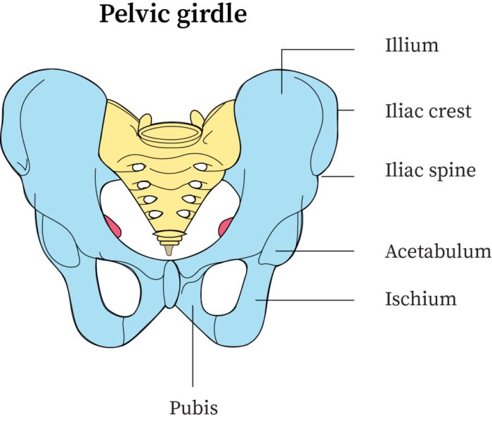
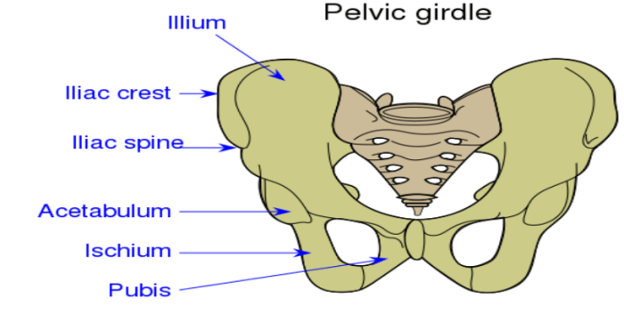
The pelvic girdle consists of three bones:
- Ilium:The largest bone of the pelvis, located at the superior and lateral aspects.
- Ischium:Situated inferiorly and posteriorly, forming the lower portion of the pelvis.
- Pubis:The most anterior bone, articulating with its counterpart to form the pubic symphysis.
| Bone | Location | Function |
|---|---|---|
| Ilium | Superior and lateral pelvis | Supports abdominal organs, attaches to lower limb |
| Ischium | Inferior and posterior pelvis | Supports weight, provides attachment for muscles |
| Pubis | Anterior pelvis | Forms pubic symphysis, provides attachment for muscles |
Bones of the Lower Limb, Art-labeling activity: bones of the pelvic girdle and lower limb
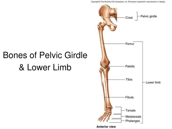
The lower limb consists of the thigh, leg, and foot, and contains the following bones:
- Femur:The longest and strongest bone in the body, forming the thigh.
- Patella:A small, triangular bone located anteriorly to the knee joint.
- Tibia:The larger and more medial bone of the lower leg.
- Fibula:The smaller and more lateral bone of the lower leg.
- Talus:The superior bone of the foot, articulating with the tibia and fibula.
- Calcaneus:The largest bone of the foot, forming the heel.
- Metatarsals:Five long bones that form the arch of the foot.
- Phalanges:Fourteen small bones that form the toes.
| Bone | Location | Function |
|---|---|---|
| Femur | Thigh | Supports weight, allows movement |
| Patella | Knee joint | Protects knee joint, allows knee extension |
| Tibia | Lower leg | Supports weight, allows movement |
| Fibula | Lower leg | Stabilizes ankle, provides attachment for muscles |
| Talus | Foot | Articulates with tibia and fibula, allows ankle movement |
| Calcaneus | Foot | Forms heel, supports weight |
| Metatarsals | Foot | Form arch of foot, allow movement |
| Phalanges | Toes | Allow toe movement |
FAQ Corner
What are the benefits of using art-labeling activities in medical education?
Art-labeling activities offer numerous benefits in medical education. They enhance visual learning, improve spatial reasoning, promote active recall, and foster a deeper understanding of complex anatomical structures.
How can I assess student learning from art-labeling activities?
To assess student learning, consider using rubrics that evaluate the accuracy and completeness of their labels, as well as their ability to identify and describe the anatomical structures.

